
The Silent Hormone Crisis: How Plastics, Food Quality, and Outdated Lab Ranges Are Failing Women's Health
A deep dive into the environmental and systemic factors driving hormonal decline in women — and why standard blood tests aren't catching it.
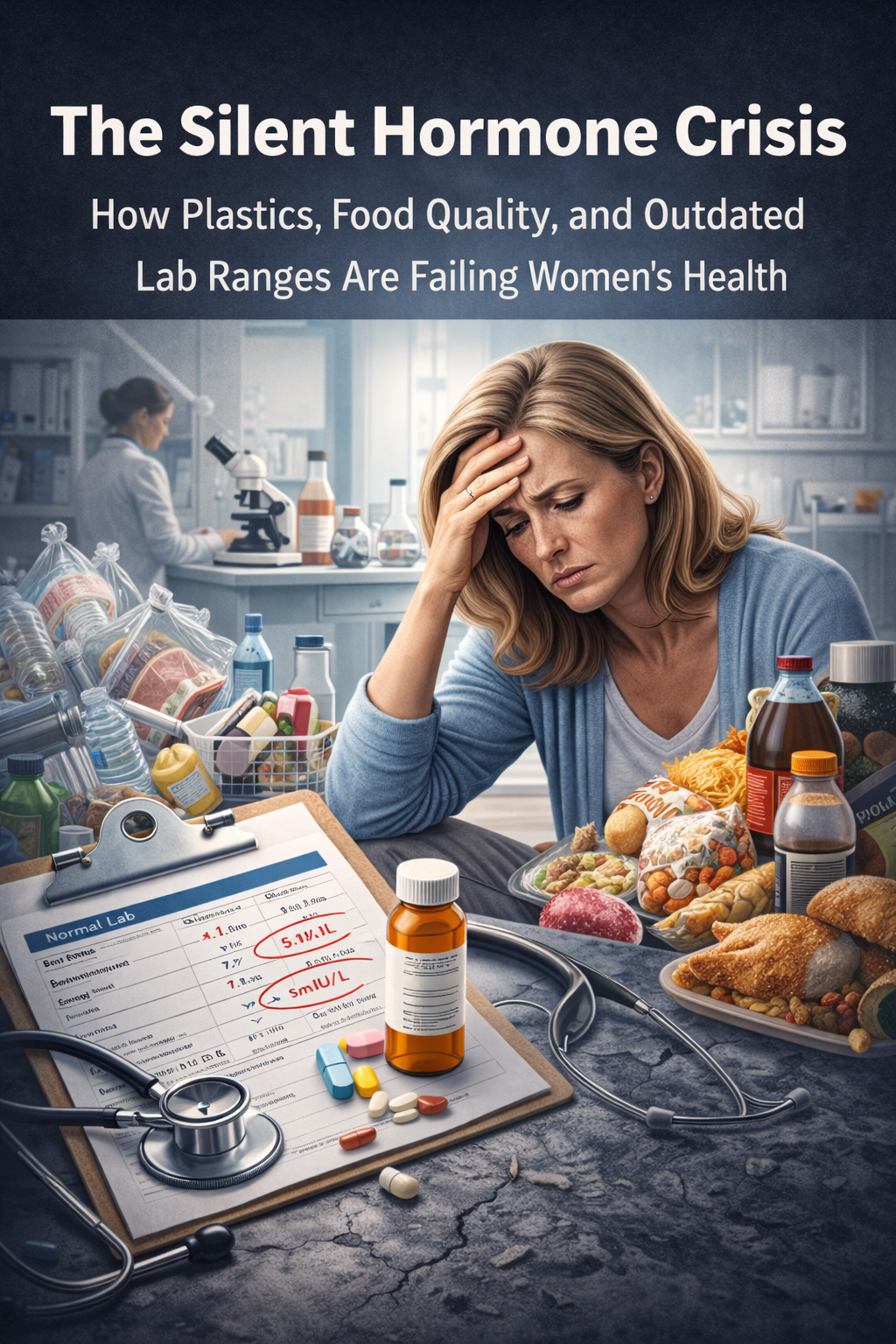
Introduction: A Perfect Storm Against Women's Hormones
Something is quietly going wrong with women's hormonal health. Rates of thyroid dysfunction, adrenal insufficiency, sleep disorders, infertility, PCOS, endometriosis, and early menopause are climbing — and the conventional medical system is struggling to explain why. The answer, increasingly supported by research, lies at the intersection of three converging crises: ubiquitous exposure to endocrine-disrupting chemicals in plastics, declining nutritional quality of our food supply, and outdated laboratory reference ranges that define "normal" based on an already-sick population.
For women, the stakes are uniquely high. Female physiology depends on intricate hormonal cascades across the thyroid, adrenal, and pineal axes — systems that are exquisitely sensitive to both toxic exposure and nutrient depletion. Yet today's standard blood tests routinely declare women "normal" while they suffer from fatigue, brain fog, weight gain, anxiety, insomnia, and reproductive dysfunction.
This blog examines the research behind each of these converging threats and makes the case for a precision medicine approach to women's hormonal health.
Part 1: The Plastic Endocrine Disruption Crisis
What Are Endocrine-Disrupting Chemicals?
Endocrine-disrupting chemicals (EDCs) are natural or synthetic substances that mimic, block, or interfere with the body's hormonal signaling. The National Institute of Environmental Health Sciences has identified over 1,000 chemicals classified as endocrine disruptors, many of them embedded in the plastics that surround us daily — from food packaging and water bottles to cosmetics, clothing, and medical devices.
The Endocrine Society's landmark 2024 report, "Endocrine Disrupting Chemicals: Threats to Human Health," raised urgent concerns about the global health threats posed by EDCs. The report found increasing evidence that these chemicals contribute to diabetes, neurological disorders, reproductive dysfunction, inflammation, and compromised immune function. Global production of plastics continues to increase even as scientists warn that chemical and plastic pollution is an escalating crisis.
The Key Offenders
Bisphenol A (BPA) and BPA Substitutes: BPA is one of the most studied EDCs. It mimics estrogen and disrupts thyroid hormone metabolism. A 2025 review published in Toxicology Mechanisms and Methods confirmed that BPA contributes to obesity, diabetes, and cardiovascular dysfunction. Its replacement, BPS, exhibits similar endocrine-disrupting properties and persists even longer in the environment. Research published in Reproductive Toxicology (2025) demonstrated that BPS exposure causes oxidative stress in ovarian tissue, disrupts folliculogenesis, and impairs the estrous cycle — effects that melatonin was shown to partially counteract.
Phthalates: These plasticizers are found in hundreds of consumer products including food packaging, cosmetics, fragrances, children's toys, and medical tubing. They are associated with reproductive issues, metabolic conditions, reduced thyroid weight during childhood exposure, and developmental abnormalities.
PFAS ("Forever Chemicals"): Per- and polyfluoroalkyl substances are used in clothing, food packaging, and non-stick coatings. Recent studies show they disrupt estrogen and testosterone signaling and impair thyroid hormone function. Their persistence in the environment and the human body makes them particularly dangerous.
Micro- and Nanoplastics (MNPs): A 2025 review in the International Journal of Molecular Sciences explored the toxicological impact of MNPs on the hypothalamus, pituitary gland, thyroid, pineal body, ovaries, and testes. These particles contain EDCs that bind to hormone receptors and disrupt the hypothalamic-pituitary-gonadal (HPG), hypothalamic-pituitary-thyroid (HPT), and hypothalamic-pituitary-adrenal (HPA) axes — the three central regulatory systems governing women's hormonal health.
Direct Impact on Women's Hormones
Thyroid disruption: Plastic additives including PBDEs, BPA, phthalates, and organotin act as thyroid-disrupting chemicals. They enter the body through the gastrointestinal tract and interfere with T4 and T3 biochemical pathways, adversely affecting thyroid hormone production and metabolism. Long-term exposure has been shown to exhaust thyroid endocrine function.
Adrenal axis disruption: MNPs disrupt the HPA axis, the central stress-response system governing cortisol production. This disruption leads to dysregulated cortisol rhythms, chronic fatigue, impaired immune function, and metabolic dysfunction — all of which disproportionately affect women.
Melatonin and pineal gland disruption: One of the most underappreciated findings is that microplastics can accumulate in the pineal gland and impair melatonin's regulatory cycle. Melatonin is not merely a "sleep hormone" — it is a potent antioxidant, a protector against mitochondrial dysfunction, a regulator of reproductive hormones, and a critical defense against the oxidative stress caused by EDC exposure. When plastic particles compromise melatonin production, they simultaneously remove the body's primary defense against the very damage they cause.
Reproductive disruption: A 2023 Rutgers study demonstrated that inhaled micro- and nanoplastic particles caused a decrease in 17-beta estradiol levels in female rats — and that it was the plastic particles themselves, not just chemical additives, driving this effect. In women, BPA exposure has been associated with hormonal imbalances, reduced ovarian reserve, PCOS, endometriosis, fibroids, and infertility. Microplastics have been found in human placentas, and their presence in women's uteruses has been linked to miscarriage.
Part 2: The Nutrient Depletion Crisis

Our Food Is Not What It Used to Be
Even women who eat a "healthy" diet are getting fewer nutrients from their food than previous generations. A landmark study by Donald Davis at the University of Texas, published in the Journal of the American College of Nutrition (2004), analyzed USDA nutritional data for 43 garden crops and found statistically reliable declines in protein, calcium, phosphorus, iron, riboflavin, and vitamin C between 1950 and 1999.
This decline has only accelerated. Key findings from multiple studies include:
Calcium levels in broccoli have decreased by more than 60% over 50 years.
Magnesium content in vegetables and wheat has declined by up to 25%.
The protein content of corn declined 30–50% from 1920 to 2001, while starch content increased.
Trace minerals including manganese, zinc, copper, and nickel have decreased in vegetable crops, while toxic minerals like aluminum, lead, and cadmium have increased.
A Kushi Institute analysis (1975–1997) found average calcium in 12 fresh vegetables dropped 27%, iron dropped 37%, vitamin A dropped 21%, and vitamin C dropped 30%.
The 1992 Earth Summit reported mineral level declines of 72% in Europe, 76% in Asia, and 85% in North America over the preceding century.
Why This Matters for Women's Hormones
The nutrients being lost from our food supply are precisely the ones required for thyroid, adrenal, and reproductive hormone production and conversion:
Selenium is essential for the deiodinase enzymes that convert T4 (inactive thyroid hormone) to T3 (the active form). Selenium deficiency directly impairs thyroid function and is associated with autoimmune thyroid disease. Soil depletion and processed food consumption have made selenium deficiency increasingly common.
Zinc plays a critical role in thyroid hormone regulation, deiodinase enzyme function, and the synthesis of both TRH and TSH. It also supports progesterone production and estrogen metabolism. Stress, hormonal contraception, and modern diets all deplete zinc.
Magnesium regulates the HPA axis and cortisol production. It supports GABA for mood stability and aids estrogen clearance. Magnesium deficiency — driven by soil depletion, chronic stress, and caffeine consumption — is linked to anxiety, insomnia, PMS, and impaired stress response. Due to modern farming practices, it is increasingly difficult to get adequate magnesium from food alone.
Iodine is the foundational building block of thyroid hormones T3 and T4. Iodine deficiency remains one of the most common causes of thyroid dysfunction worldwide.
Iron and ferritin are essential for thyroid hormone production. Functional practitioners note that ferritin levels below 50 ng/mL are often symptomatic, yet many standard lab ranges set the lower limit far below this threshold.
Vitamin D has immune-modulating properties and correlates inversely with thyroid antibodies — higher vitamin D levels are associated with lower autoimmune thyroid markers.
B vitamins (B2, B3, B6, B12) are critical cofactors for thyroid hormone conversion, neurotransmitter production, and adrenal function.
The Compounding Effect
As one clinical review noted: "People today experiencing constant and multiple stressors have fewer nutrients to replenish challenged adrenals." The nutrients consumed in greatly increased quantities during high-stress states are not being replenished by the modern diet. Even in "healthy" foods, nutrient quality has been diminished by chemical fertilizers that deplete the soil of the minerals necessary for plants to produce the vitamin- and phytonutrient-rich foods that previously existed.
Women are caught in a double bind: their hormonal systems require more nutritional support than ever due to chronic stress and toxic exposure, yet the food supply delivers less and less of what they need.
Part 3: The Reference Range Problem — Why Standard Blood Tests Are Missing the Crisis
How "Normal" Ranges Are Determined
Standard laboratory reference ranges are determined by measuring values in a population of tested individuals and defining the middle 95% as "normal." The 2.5% above and below are flagged as abnormal. This statistical approach has a critical flaw: the reference population includes people who are already experiencing subclinical dysfunction.
As functional medicine practitioners have long argued, if you measure thyroid hormones in a population where a significant percentage already has undiagnosed thyroid problems, you create a "normal" range that encompasses dysfunction. The result: women with clear hypothyroid symptoms are told their labs are "normal."
The TSH Controversy
The conventional reference range for TSH (thyroid stimulating hormone) is typically 0.4–4.0 or even up to 5.0 mIU/L. However:
The National Academy of Clinical Biochemistry found that more than 95% of normal individuals (those without thyroid disease) have a TSH below 2.5 mIU/L.
The American Association of Clinical Endocrinologists recommended narrowing the TSH range as far back as 2003, after it was discovered that people with existing thyroid dysfunction had been included in the original reference population.
A 2024 study from the Thyroid Studies Collaboration, published after analyzing over 134,000 participants, found that individuals with Free T4 levels in the 20th–40th percentile had the least risk of death and cardiovascular disease. Those in the 80th–100th percentile had a 57% higher risk of heart disease-related death and 34% higher risk of all-cause mortality — even within the "normal" range.
A 2025 study published in the Annals of Internal Medicine found that using age-, sex-, and race-specific reference intervals could reclassify many thyroid diagnoses, with nearly half of subclinical hypothyroid cases potentially being over- or mis-diagnosed under current ranges.
In functional and precision medicine, the optimal TSH range is generally considered 0.5–2.0 mIU/L, with most women feeling their best when TSH falls between 1.0 and 2.0, Free T4 is above 1.1 ng/dL, and Free T3 is above 3.2 pg/mL.
Beyond TSH: What's Being Missed
Standard thyroid panels typically measure only TSH and sometimes Free T4. This misses critical aspects of thyroid physiology:
Free T3 — the active hormone that actually powers metabolism, cognition, and energy. A woman can have "normal" TSH and T4 while having critically low T3 due to conversion problems.
Reverse T3 — produced when the body is under stress, effectively blocking T3 at the receptor. Elevated Reverse T3 with normal TSH creates a functionally hypothyroid state invisible to standard testing.
TPO and Thyroglobulin Antibodies — markers of autoimmune thyroid disease (Hashimoto's), which can be present and causing damage long before TSH becomes abnormal.
Nutrient cofactors — selenium, zinc, ferritin, vitamin D, and B vitamins that are essential for thyroid function but are almost never tested alongside thyroid panels.
Adrenal Hormones: Cortisol and DHEA
The standard lab reference range for morning cortisol is typically 10–20 mcg/dL. Functional medicine practitioners prefer a range of 15–20 mcg/dL, recognizing that lower values — while "normal" by lab standards — are often associated with significant symptoms of HPA axis dysregulation.
More importantly, a single morning cortisol blood draw provides only a snapshot. The cortisol rhythm is diurnal, and dysfunction often manifests as pattern disruption: elevated cortisol at night (causing insomnia), flattened rhythms (causing exhaustion), or mismatched cortisol-to-DHEA ratios. Multi-point salivary cortisol testing provides far more clinical insight, yet it remains outside standard care.
Melatonin: The Forgotten Hormone
Melatonin secretion progressively declines with age, and strong reductions are observed in Alzheimer's disease, cardiovascular disease, cancer, diabetes, and numerous other conditions. The pineal gland has the highest calcification rate of any organ in the human body, and this calcification directly jeopardizes melatonin's synthetic capacity.
Yet melatonin is virtually never tested in standard blood work. In the context of widespread plastic exposure — which has been shown to impair pineal function and melatonin's regulatory cycle — this omission is particularly consequential. Melatonin is not only essential for sleep; it supports ovarian function, mitigates oxidative stress, protects against BPA-induced gut damage, promotes testosterone and estrogen synthesis, and acts as a critical antioxidant throughout the body. Research has shown that melatonin receptors are present in reproductive organs, and melatonin levels in ovarian follicular fluid exceed those in serum and increase with follicular development.
Part 4: Why Women Need Higher — Not Lower — Hormonal Thresholds
The Unique Demands of Female Physiology
Women's bodies cycle through hormonal fluctuations on daily, monthly, and lifetime scales that demand robust thyroid, adrenal, and pineal function. Consider:
The thyroid regulates basal metabolic rate, body temperature, heart rate, digestion, fertility, and cognitive function. Women are 5–8 times more likely than men to develop thyroid dysfunction, with 1 in 8 women experiencing thyroid problems in their lifetime.
The adrenals manage the stress response, blood sugar regulation, immune function, and sex hormone production (particularly important as ovarian function declines in perimenopause and menopause). Women in modern society face chronic, unrelenting stress that demands sustained cortisol output.
The pineal gland and melatonin govern circadian rhythm, sleep architecture, antioxidant defense, reproductive timing, and immune surveillance — all of which are essential for women navigating hormonal transitions from menarche through menopause.
When blood tests use broad reference ranges that define the bottom of "normal" at levels associated with symptomatic dysfunction, women are left without diagnosis or treatment during the very periods when their bodies need optimal hormonal support the most.
The Precision Medicine Imperative
A precision medicine approach to women's hormonal health would include:
Comprehensive thyroid panels — TSH, Free T4, Free T3, Reverse T3, TPO antibodies, and thyroglobulin antibodies, interpreted against functional (not just statistical) ranges.
Multi-point cortisol and DHEA testing — salivary or dried urine testing that maps the diurnal cortisol curve rather than relying on a single blood draw.
Melatonin assessment — particularly in women with sleep disruption, fertility challenges, or high toxic exposure.
Nutrient cofactor panels — selenium, zinc, magnesium, ferritin, vitamin D, iodine, and B vitamins, with functional thresholds (not just "absence of deficiency" cutoffs).
Environmental exposure assessment — evaluation of BPA, phthalate, and PFAS body burden in women with unexplained hormonal dysfunction.
Part 5: What Women Can Do Now
Reduce Exposure
Replace plastic food storage with glass or stainless steel.
Avoid heating food in plastic containers or with plastic wrap.
Choose personal care products free of phthalates, parabens, and synthetic fragrances.
Filter drinking water to remove PFAS and other contaminants.
Choose organic produce when possible to reduce pesticide and glyphosate exposure.
Avoid canned foods and beverages lined with BPA-containing epoxy resins.
Restore Nutrient Foundations
Prioritize nutrient-dense, whole foods — leafy greens, wild-caught fish, pastured eggs, organ meats, nuts, seeds, and fermented foods.
Support thyroid function with selenium-rich foods (Brazil nuts, sardines), zinc (pumpkin seeds, oysters), and iodine (seaweed, fish).
Address magnesium depletion through supplementation and magnesium-rich foods (dark leafy greens, dark chocolate, avocados).
Consider working with a practitioner to assess and optimize ferritin, vitamin D, and B vitamin status.
Buy from local, organic, and regeneratively farmed sources whenever possible.
Advocate for Better Testing
Request comprehensive thyroid panels, not just TSH.
Ask for cortisol pattern testing (4-point salivary or DUTCH testing).
Find a practitioner who uses functional reference ranges and treats the whole clinical picture — not just a number on a lab report.
Trust your symptoms. A "normal" lab result does not mean you are functioning optimally.
Conclusion: Redefining "Normal" for Women's Health
We are living in an unprecedented era of environmental hormonal assault. Plastics and their chemical additives disrupt every major endocrine axis. Our food supply delivers fewer of the nutrients women's bodies need to produce, convert, and utilize hormones. And the laboratory standards designed to catch disease were never calibrated to identify optimal function — particularly in women.
The convergence of these three factors creates a silent epidemic: women whose hormonal systems are being degraded by toxic exposure and nutrient depletion, but whose standard blood work shows "normal" results because the reference ranges themselves reflect a population that is already compromised.
Precision medicine, functional testing, environmental awareness, and a return to nutrient-dense food are not luxury approaches — they are urgent necessities. Women deserve laboratory ranges that reflect thriving, not merely the absence of overt disease. They deserve practitioners who test comprehensively and listen to symptoms rather than dismissing them based on outdated statistical averages.
The hormone crisis is real. The science is clear. And the solutions are within reach — if we are willing to look beyond "normal."
References and Key Sources
Endocrine Society (2024). Endocrine Disrupting Chemicals: Threats to Human Health. Co-produced with IPEN.
Toxicology Mechanisms and Methods (2025). "Implications of plastic-derived endocrine disruptors on human health." Vol 35(8): 894-918.
International Journal of Molecular Sciences (2025). "Micro- and Nanoplastics as Disruptors of the Endocrine System." MDPI.
National Institute of Environmental Health Sciences (NIEHS). "Endocrine Disruptors" — health topics overview.
Rutgers University (2023). "Plastic particles themselves, not just chemical additives, can alter sex hormones." ScienceDaily.
ScienceDirect (2025). "Involvement of autophagy and mitophagy modulation in the protective effect of melatonin against BPS-induced ovarian toxicity." Reproductive Toxicology.
Frontiers in Cell and Developmental Biology (2024). "Melatonin alleviates oxidative stress damage in mouse testes induced by bisphenol A."
ScienceDirect (2025). "Pineal melatonin improves bisphenol A-induced enterotoxicity via tryptophan/AhR/mitochondria signaling in pregnant mice."
PMC (2023). "A review of the endocrine disrupting effects of micro and nano plastic and their associated chemicals in mammals."
American Thyroid Association (2024). "The 'new normal' for thyroid function test ranges." Clinical Thyroidology for the Public.
Thyroid Studies Collaboration (2023). "The optimal healthy ranges of thyroid function defined by the risk of cardiovascular disease and mortality."
Annals of Internal Medicine (2025). "Using age, sex, and race-specific standards could reclassify many thyroid disease diagnoses."
National Academy of Clinical Biochemistry. Data indicating 95% of normal individuals have TSH below 2.5 mIU/L.
Davis, D. et al. (2004). "Changes in USDA Food Composition Data for 43 Garden Crops, 1950 to 1999." Journal of the American College of Nutrition.
Scientific American. "Dirt Poor: Have Fruits and Vegetables Become Less Nutritious?"
PMC (2024). "An Alarming Decline in the Nutritional Quality of Foods: The Biggest Challenge for Future Generations' Health."
PMC (2012). "Melatonin in Aging and Disease — Multiple Consequences of Reduced Secretion."
MDPI Molecules (2018). "Pineal Calcification, Melatonin Production, Aging, Associated Health Consequences and Rejuvenation of the Pineal Gland."
Wilson, J.L. (2014). "Clinical perspective on stress, cortisol and adrenal fatigue." Complementary Therapies in Clinical Practice.
ScienceDirect (2023). "Endocrine disruptors: Unravelling the link between chemical exposure and Women's reproductive health."